
Cancer Screening :
Cancer screening aims to detect cancer before symptoms appear. This may involve blood tests, urine tests, other tests, or medical imaging. The benefits of screening in terms of cancer prevention, early detection and subsequent treatment must be weighed against any harms.
Screening tests must be effective, safe, well-tolerated with acceptably low rates of false positive and false negative results.
Screening for cancer can lead to cancer prevention and earlier diagnosis. Early diagnosis may lead to higher rates of successful treatment and extended life. However, it may also falsely appear to increase the time to death through lead time bias or length time bias.
1. Breast Cancer Screening :
Breast cancer screening is the medical screening of asymptomatic, apparently healthy women for breast cancer in an attempt to achieve an earlier diagnosis.The assumption is that early detection will improve outcomes. A number of screening tests have been employed, including clinical and self breast exams, mammography, genetic screening, ultrasound, and magnetic resonance imaging.
There are mainly Three types of tests used for Breast Cancer Screening as :
1.1 Breast Self Examination (BSE) :
Breast self-examination (BSE) is a screening method used in an attempt to detect early breast cancer. The method involves the woman herself looking at and feeling each breast for possible lumps, distortions or swelling.
Opportunity for woman to become familiar with her breasts
Monthly exam of the breasts and underarm area
May discover any changes early
Begin at age 20, continue monthly
Breast Self Exam Information :
It is easy to do and the more you do it, the better you will get at it.When you get to know how your breasts normally feel, you will quickly be able to feel any change, and early detection is the key to successful treatment.Most breast lumps are found by women themselves, but in fact, most lumps in the breast are not cancer.The best time to do breast self-exam is right after your period, when breasts are not tender or swollen. If you do not have regular periods or sometimes skip a month, do it on the same day every month.When to do a self Breast Exam : The best time to do breast self-exam is right after your period, when breasts are not tender or swollen. If you do not have regular periods or sometimes skip a month, do it on the same day every month.
There are two parts to the BSE :
1. Looking
2. Feeling
Looking :
Looking in Mirror

Lump

Change in Skin color, Size

Skin Dimpling

Changes in Nipple

Nipple Discharge

Lump :
- Any new lump or hard knot found in the breast or armpit.
- Any new lump or thickening that does not shrink or lessen after your next period.
Change in Skin color, Size :
- Any change in size, shape or symmetry of your breast
- Any thickening or swelling of the breast
Skin Dimpling :
- Any dimpling, puckering or indention in the breast
- Dimpling, skin irritation or other change in the breast skin or nipple
Changes in Nipple :
- Redness or scaliness of the nipple or breast skin
- Nipple tenderness or pain
- Nipple retraction; turning or drawing inward or pointing in a new direction
Nipple Discharge :
- Any fluid coming from your nipples other than breast milk, particularly if the discharge is bloody, clear and sticky, dark or occurs without squeezing your nipple
Feeling :
- Examine each breast separately.
- Use pads of fingers not the tips.
- Examine the armpits.
Finger Use :
- Your finger pads are the top third of each finger, not the tips.
- Use the pads of your middle three fingers to feel the texture of your breast.
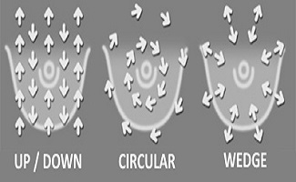
Patterns :
- Up / Down
- Circular
- Wedge
When using any of the 3 patterns, you should always be using a circular rubbing motion (in dime-sized circles) without lifting up your fingers.

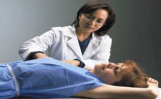
1.2. Clinical breast examination (CBE) :
A Clinical Breast Examination (CBE) is a physical examination of the breast done by a health professional. Clinical breast examinations are used along with mammograms to check women for breast cancer.
A Clinical Breast Examination may be part of your regular checkup. Talk with your health professional about how often you need a breast examination. Women with breast implants should also have regular clinical breast examinations.
- Performed by doctor or trained nurse practitioner
- Annually for women over 40
- At least every 3 years for women between 20 and 40
- More frequent examination for high risk patients
The Procedure :
- Explain what you will be doing
- Ask if she does breast self exam
- Warm your hands
- Assure privacy
- Would someone else in the room be helpful?
- Assist patient to supine position
Palpation :
Variables important in palpating the breast correctly are :
- patient position.
- breast boundaries.
- examination pattern.
- finger position, movement, and pressure.
- duration of the examinatio.

1. Patient Position :
Patient’s Positions required during examination are :
Clinical breast examination requires flattening breast tissue against the patient’s chest
Patient is supine during the examination
2. Breast Boundaries :
- Breast tissue extends laterally toward the axilla and superiorly toward the clavicle
- Cover a rectangular area bordered by the clavicle superiorly, the midsternum medially, the midaxillary line laterally, and line passing through xiphisternum inferiorly
4. Technique :
- The 3 middle fingers are held together, with the metacarpal-phalangeal joint slightly flexed
- Pads of the fingers are the examining surface
- Each area is palpated by making small circles using 3 different pressures-light, medium, and deep
3. Examination Pattern :
- Palpation begins in the axilla and extends in a straight line down the midaxillary line to the inferior axis line
- The fingers move medially, and palpation continues up the chest in a straight line to the clavicle
- Rows should be overlapping
5. Duration :
- 3 minutes recommended for each breast
- Average actual time spent is 1.8 minutes
- Discuss with patients the time needed to do a complete examination and discuss the procedure during the examination
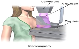
1.3. Mammography :
Mammography is the process of using low-energy X-rays (usually around 30 kVp) to examine the human breast, which is used as a diagnostic and screening tool. The goal of mammography is the early detection of breast cancer, typically through detection of characteristic masses and/or microcalcifications.
- A screening mammogram is an x-ray examination of the breast in a woman who has no symptoms.
- The goal of a screening mammogram is to find cancer when it is still too small to be felt by a woman or her doctor.
- Effective in women above age of 50 yrs.
- Based on fair evidence, screening mammography in women aged 40 to 70 years decreases breast cancer mortality.
- The benefit is higher for older women, in part because their breast cancer risk is higher.
- Women (asymptomatic) 40 years of age and older should have a mammogram every year.
2. Cervical Cancer Screening :
Cervical screening is a way of preventing cancer by finding and treating early changes in the neck of the womb (cervix). These changes could lead to cancer if left untreated. The screening uses a test called cytology, which many people know as the smear test.
A nurse or doctor takes a sample of cells from the cervix with a small brush. They send the sample to a laboratory to be checked for abnormalities. In some cases, samples are also tested for a virus called human papilloma virus (HPV) that increases the risk of cervical cancer.
There are two types of tests used for cervical cancer screening as :
1. Pap Test :
The Pap test can find early cell changes and treat them before they become cancer. The Pap test can also find cervical cancer early, when it’s easier to treat.
The pap test, known earlier as Pap smear, cervical smear, or smear test is a method of cervical screening used to detect potentially pre-cancerous and cancerous processes in the endocervical canal (transformation zone) of the female reproductive system.
A Pap smear is performed by opening the vaginal canal with a speculum, then collecting cells from the outer opening of the cervix of the uterus and the endocervix. The cells are examined under a microscope to look for abnormalities. The test aims to detect potentially pre-cancerous changes (called cervical intraepithelial neoplasia (CIN) or cervical dysplasia), which are caused by sexually transmitted human papillomaviruses. The test remains an effective, widely used method for early detection of pre-cancer and cervical cancer. The test may also detect infections and abnormalities in the endocervix and endometrium.
2. HPV (Human Papilloma Virus) Test :
The HPV (Human Papilloma Virus) test finds certain infections that can lead to cell changes and cancer. HPV infections are very common, and most go away by themselves and don’t cause these problems. The HPV test may be used along with a Pap test, or to help doctors decide how to treat women who have an abnormal Pap test.
The human papillomavirus (HPV) test detects the presence of human papillomavirus, a virus that can lead to the development of genital warts, abnormal cervical cells or cervical cancer.
Your doctor might recommend the HPV test if:
- Your Pap test was abnormal, showing a typical squamous cells of undetermined significance (ASCUS)
- You’re age 30 or older
The HPV test is available only to women; no HPV test yet exists to detect the virus in men. However, men can be infected with HPV and pass the virus along to their sex partners.
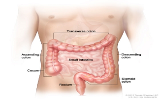
3. Colon Cancer Screening :
Colorectal cancer (also known as colon cancer, rectal cancer or bowel cancer) is the development of cancer in the colon or rectum. It is due to the abnormal growth of cells that have the ability to invade or spread to other parts of the body.
Signs and symptoms :
-
- blood in the stool
- change in bowel movements
- weight loss
- feeling tired all the tim
smoking, and not enough physical activity. Dietary factors that increase the risk include red and processed meat, as well as alcohol. Another risk factor is inflammatory bowel disease, which includes Crohn’s disease and ulcerative colitis.
Colon cancer screening can detect polyps and early cancers in the intestines. This type of screening can find problems that can be treated before cancer develops or spreads. Regular screenings may reduce the risk of death and pain caused by colorectal cancer.
There are There are several ways used for colon cancer screening as :
- Stool Test :Polyps in the colon and smaller cancers can cause small amounts of bleeding that cannot be seen with the naked eye. But the blood can often be found in the stool.This method checks your stool for blood.The most common test used is the fecal occult blood test (FOBT). Two other tests are called the fecal immunochemical test (FIT) and stool DNA test (sDNA).
- Sigmoidoscopy :This test uses a flexible small scope to view the lower part of your colon. Because the test only looks at the last one-third of the large intestine (colon), it may miss some cancers that are higher in the large intestine.Sigmoidoscopy and a stool test should be used together.
- Colonoscopy :A colonoscopy is similar to a sigmoidoscopy, but the entire colon can be viewed.During a colonoscopy, you receive medicine to make you relaxed and sleepy.Sometimes, CT scans are used as an alternative to a regular colonoscopy. This is called a virtual colonoscopy.
- Other Tests :Double-contrast barium enema is a special x-ray of the large intestine that looks at the colon and rectum.Capsule endoscopy involves swallowing a small, pill-sized camera.
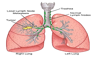
4. Lung Cancer Screening :
Lung cancer, also known as carcinoma of the lung or pulmonary carcinoma, is a malignant lung tumor characterized by uncontrolled cell growth in tissues of the lung.
If Lung Cancer left untreated, this growth can spread beyond the lung by process of metastasis into nearby tissue or other parts of the body. Most cancers that start in the lung, known as primary lung cancers, are carcinomas that derive from epithelial cells. The main primary types are small-cell lung carcinoma (SCLC) and non-small-cell lung carcinoma (NSCLC).
Signs and symptoms :
-
-
-
- respiratory symptoms : coughing, coughing up blood, wheezing or shortness of breath.
- systemic symptoms : weight loss, fever, clubbing of the fingernails, or fatigue.
- symptoms due to the cancer mass pressing on adjacent structures : chest pain, bone pain, superior vena cava obstruction, difficulty swallowing.Lung cancer screening refers to cancer screening strategies used to identify early lung cancers before they cause symptoms,at a point where they are more likely to be curable. Screening studies for lung cancer have only been done in high risk populations, such as smokers and workers with occupational exposure to certain substances.
-
-
The tests used for Lung cancer screening is :
1. Low-Dose Computed Tomography ( CT scan, LDCT) :
-
-
-
- In this test, an X-ray machine scans the body and uses low doses of radiation to make detailed pictures of the lungs Test .
-
-
2. Chest x-ray :
-
-
-
- Many healthcare providers recommend an annual chest x-ray for patients who smoke.
- In a large study comparing chest x-ray to CT for lung cancer screening, only CT showed reduced risk of death.
- Current guidelines recommend against screening at-risk subjects by chest x-ray.
-
-
3. Sputum Tests :
-
-
-
- Analyzing a patient’s sputum for evidence of cancer cells in order to detect lung cancer.
- So far, no clear benefit to this approach has been found. Additional studies that use new technologies to examine the sputum are underway.
-
-
4. PET scan :
-
-
-
- Positron Emission Tomography (or PET scanning, which uses a small amount of radioactivity to provide a detailed picture of an organ’s function) has been used in combination with CT scanning (PET/CT).
- It involves a higher dose of radiation than CT alone and benefit has not been shown for screening purposes.
-
-
5. Other studies :
-
-
-
- Direct visualization of the lungs with bronchoscopy and breath analysis for cancer markers are two tests.
-
-